Advanced Chemicals and Materials Analysis Service (ACMA)

Description
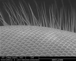
Electron Microscopy
Scanning electron microscopy (SEM) enables imaging of a surface using an electron beam. The electron beam interacts with the surface of the sample and by using a variety of detectors topographic images, atomic-number (Z) contrast images, element composition and fluorescence information can be obtained from the specimen.
In our microscopy suite we have two SEMs and an ESEM which provide a range of analytical techniques. Our capabilities include:
•Digital imaging over a range of magnifications depending on sample quality (~ X20 to X300 000), in the following modes ◦secondary electron emission (SEI) - ideal for materials with surface relief and/or multiple faces ◦backscattered electron (BSE) - best suited to thin section work ◦cathodoluminescence (CL) - samples must contain luminescent elements and be thin section form
•Energy Dispersive X-ray Spectroscopy (EDS or EDX) ◦EDX detectors allow the ID and semi-quantification of elements from carbon (C) upwards ◦output is in the form of spot/point data, average area measurements or single element digi-mapping (using X-ray intensity distribution)
•Low vacuum (LV) mode for sensitive samples or those not appropriate for carbon or gold coating (typically used to improve conductivity)
•Electron backscatter diffraction EBSD (only on the XL30 ESEM)
Samples
•Your samples should all be clearly labelled with unique identifiers •Dry powder and material samples are usually best stored in glass containers and not in plastic bags or bottles (as plastics are prone to static and some can contaminate samples) •Disposal - we will dispose of all unclaimed samples after three months from analysis completion
X-ray Powder Diffraction
XRPD is a robust technique used for the characterisation of crystalline materials, i.e. those which have a degree of order to their structure. By exposing a sample to an X-ray beam of known wavelength, a diffraction pattern unique to each crystalline phase within that sample material is generated and this pattern is used to identify and characterise each crystalline phase present.
Through interpretation of diffraction patterns, XRPD analysis allows:
•identification of crystalline phases •measurement of crystallite/crystal domain size within discrete crystalline phases •calculation of unit cell dimensions through pattern indexing •phase quanitification using one of several methods The PANalytical X'Pert Pro Multipurpose Diffractometer (MPD) is used for most of our X-ray powder diffraction (XRPD) analysis. It has various interchangeable attachments:
•X'Celerator (RTMS detector) +/- monochromator •Anton Paar HTK2000 heating stage •automated sample changer (15-berth magazine) •sample spinner stage or sample bracket options
We also have a PW1730 X-ray generator with a sample spinner stage.
Although our XRPD lab is primarily a powder diffraction facility, we often carry out phase identification in thin film samples. Contact us if you are interested in thin film analysis - we will try to accommodate your analytical requests.
Complimentary to our XRPD lab is the X-ray crystallography laboratory in the School of Chemistry who have extensive expertise in crystal structure determinations using single-crystal diffractometers.
Chemical Analysis
In our chemistry section we provide a range of analytical techniques:
•Inductively Coupled Plasma Optical Emission Spectroscopy (ICP-OES) •Fourier Transform Infrared Spectroscopy (FTIR) •Carbon, Hydrogen and Nitrogen Combustion Analysis (CHN)
Manufacturer
Model
Contact
Academic Contact
| Dr Alasdair Charles | |
| alasdair.charles@ncl.ac.uk |
Technical Contact
| ACMA Facility Email | |
| acma@ncl.ac.uk |