Scanning Electron Microscope Tescan

Description
Please contact Newcastle University's Electron Microscopy Research Services to arrange bookings on this equipment, or to find out about services offered by the facility.
www.ncl.ac.uk/emrs
em.researchservices@ncl.ac.uk
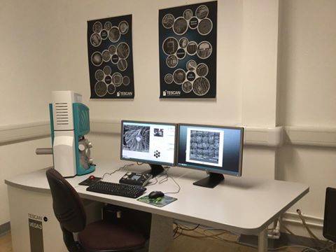
Scanning electron microscope for the visualisation of biological and material samples. Sample preparation is also available (critical point dryer and gold-coater).
A scanning electron microscope (SEM) is a type of electron microscope that produces images of a sample by scanning it with a focused beam of electrons. The electrons interact with atoms in the sample, producing various signals that can be detected and that contain information about the sample's surface topography and composition. The electron beam is generally scanned in a raster scan pattern, and the beam's position is combined with the detected signal to produce an image. SEM can achieve resolution better than 1 nanometer. Specimens can be observed in high vacuum, in low vacuum, in wet conditions (in environmental SEM), and at a wide range of cryogenic or elevated temperatures.
The most common mode of detection is by secondary electrons emitted by atoms excited by the electron beam. On a flat surface, the plume of secondary electrons is mostly contained by the sample, but on a tilted surface, the plume is partially exposed and more electrons are emitted. By scanning the sample and detecting the secondary electrons, an image displaying the topography of the surface is created.
ManufacturerTescan
ModelVega LMU
Contact
Academic Contact
| Dr Kathryn White | |
| kathryn.white@ncl.ac.uk |
Technical Contact
| Mrs Tracey Davey | |
| tracey.davey@ncl.ac.uk |